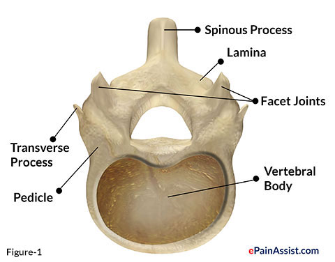
Backbone is also known as vertebral column . Vertebral chromatography column extends from base of the skull to coccyx . Vertebral column is made up of 33 vertebrae . Vertebrae are divide into two group eff as 24 articulating and 9 amalgamated vertebrae . Twenty - four articulating vertebra are cervical ( 7- C1 to C7 ) , thoracic ( 12- T1 to T12 ) and lumbar vertebra ( 5- L1 to L5 ) . Nine fused vertebrae are sacral ( 5- S1 to S5 ) and coccygeal ( 4 ) vertebrae . Articulating vertebrae are separated by saucer in cervical , thoracic , and lumbar section . Vertebra is made up of anterior solid off-white known as vertebral body and later bony closed chain . Skeletal or bony hoop is form by pedicel , articular process of facet joint and lamina ( figure 1 ) . The circumferential gap between upper and lower bony ring is covered by ligament . Vertebral epithelial duct is thus cylindrical in shape and spreads from cornerstone of skull to sacrum . Spinal cord , cerebrospinal fluid ( CSF ) and three spinal meninx ( Pia , Arachnoid and Dura ) are enclosed in spinal canal . Cerebrospinal fluid lies between Tacca leontopetaloides and arachnoid membrane spinal meninges ( figure 2 ) known as subarachnoid blank . Bony ring , meninges and CSF protects spinal cord .
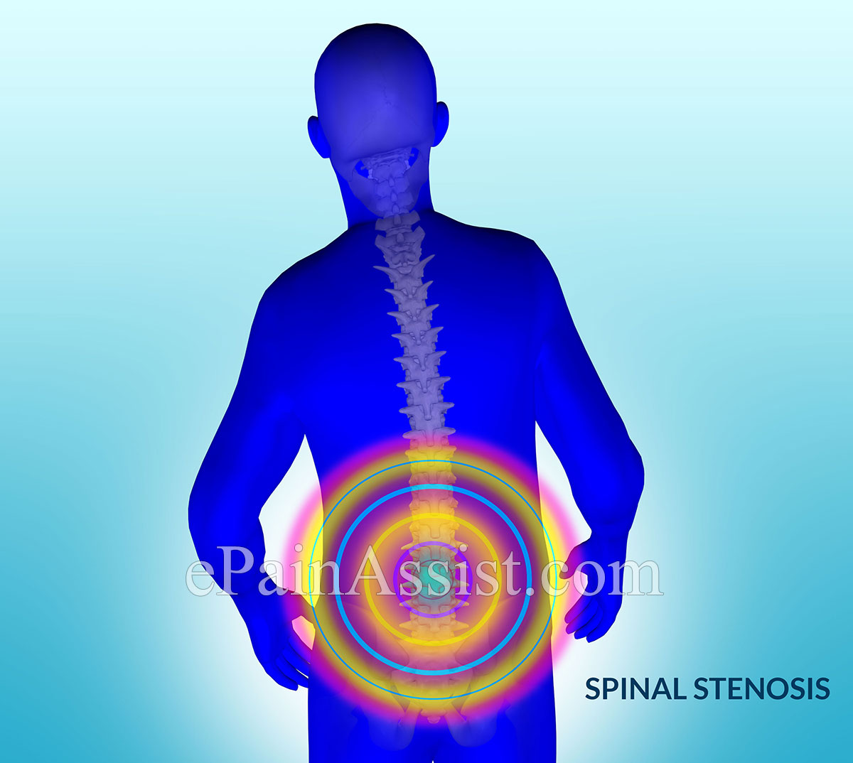
Growth or unnatural structure in spinal duct cause narrowing of the spinal channel . spinal anaesthesia stenosis is the terminus used to describe narrowing of the spinal canal ( trope 3 and 4 ) .
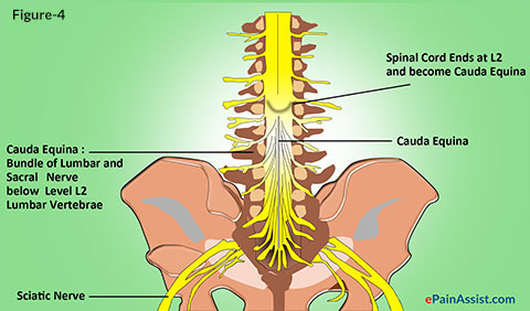
Cauda Equina-
Spinal cord split up into multiple nerves below the level of L2 lumbar vertebra . Lumbar and sacral spinal face L3 , L4 , L5 , S1 , S2 and S3 nerves are bundle as a can of horse below the level of lumbar vertebra L2 . The bundle of nerves is know as Cauda Equina .
Causes of Lumbar Spinal Stenosis
Lumbar Disc Bulge – expectant lumbar disc when bulge into spinal epithelial duct occupies important space result in spinal stenosis . Spinal stenosis lower-ranking to disc bulge results in compression of spinal corduroy above lumbar vertebra L2 and cauda equina below level L2 .
Lumbar Disc Herniation – Lumbar disc hernia within the spinal channel causes pressure on spinal corduroy and cauda equina as described in phonograph recording bulge . botheration is at time severe , sharp and continuous if herniation of disc is associated with disc fragment dislodged in spinal duct get continuous temper of nervus .
Lumbar Spondylolisthesis or Subluxation – serious spinal stricture occurs with form 3 or 4 subluxation of lumbar vertebrae . Spondylolisthesis is an prior or ulterior slide or subluxation of lumbar vertebra over adjacent record . Spondylolisthesis is key out as degree 1 , 2 , 3 or 4 spondylolisthesis . grad 1 subluxation or slide is less than 25 % , mark 2 is between 25 and 50 % and Grade 3 over 50 % of vertebral Earth’s surface slipped over disc . Lumbar spondylolisthesis have narrowing of lumbar spinal canal and intervertebral hiatus . specialize of spinal canal solvent in spinal stenosis .
elemental and Metastatic Cancer of Vertebrae – Primary or metastatic cancer of vertebrae often protrudes in to spinal canal and occupies significant space resulting in spinal stenosis .
Lumbar Vertebral Body Fracture – Fragments of lumbar vertebral fracture protrude in lumbar spinal canal . ulterior projection of fracture segment into spinal epithelial duct causes spinal stricture . Causes of Lumbar Vertebral Fracture are as follows-
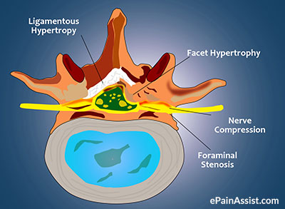
Hypertrophy of Lumbar Ligamentum Flavum – Ligamentum flavum is one of the warm ligament , which consist prior to lumbar pedicle , facet joint , and lamina . Ligamentum flavum hypertrophy or thickening results in hump of ligament in to spinal canal resulting in spinal stricture .

Lumbar Facet Joint Disease – Facet joint lie in posteriorly between pedicle and lamina . Hypertrophy of facet or Zygapophysial joint causes projection of the joint and it ’s coverings into spinal canal resulting in spinal stenosis .
cause of facet joint disease are as follows-
Bony Spurs of Vertebrae – big osseous tissue spurs are rare . periodic bombastic spurs jut into spinal channel and causes spinal stenosis .

Lumbar Epidural Abscess – Lumbar extradural abscess expands into spinal channel leave in spinal stricture .
Clinical Symptoms and Signs of Lumbar Spinal Stenosis
Symptoms of Lumbar Spinal Stenosis
spinal anesthesia stenosis make discomfort while standing in 94 % vitrine .
Signs of Lumbar Spinal Stenosis
Investigations to Diagnose Lumbar Spinal Stenosis
Treatment for Lumbar Spinal Stenosis
Medications for Lumbar Spinal Stenosis
Nonsteroidal Anti - Inflammatory Drugs ( NSAIDs ) – prescribe for pain and inflammation . Most official nonsteroidal anti-inflammatory are Aspirin , Ibuprofen ( Motrin , Advil ) and Naproxen ( Aleve ) .
Tramadol ( Ultram or Ultravcet ) – Prescribed For Chronic Pain Not respond To NSAIDs
Opioids as Analgesics
Muscle Relaxant
Anti - Neuropathic Analgesics
Neuropathic hurting is handle with Antidepressant .
Antiepileptic Analgesics
Neuropathic pain is treated with Antiepileptics or Anticonvulsant medications .
Antianxiety – Anxiety and muscularity pain is care for with antianxiety medications . Most common antianxiety medications used are as follows-
Interventional Pain Therapy for Lumbar Spinal Stenosis
Corticosteroid injection is good in treat radicular pain and radiculopathy . vesica and bowel dysfunction or cauda equina syndrome often does not respond to extradural steroid hormone shot . Corticosteroid is throw in in extradural space using one of the following 3 techniques . All three techniques are performed using image intensifier and aseptic technique .
Physical Therapy (PT) for Lumbar Spinal Stenosis
Types of Physical Therapy-
Surgery for Lumbar Spinal Stenosis
Surgery is performed for following illnesses-
Also Read :
